いろいろ e coli microscope 1000x 277892-How to identify e coli under a microscope
The primary habitat of E coli is in the gastrointestinal (GI) tract of humans and many other warmblooded animalsOn Endo agar it looks like lactose negative)All four strains are mannitol positive (best seen in fig D), cellobiose negative (strains A, B)4 Applying counterstain (safrinin) to bacterial smear as last step of endospore stain;

Bacteria Under The Microscope E Coli And S Aureus Youtube
How to identify e coli under a microscope
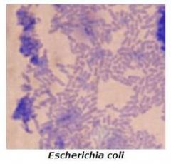
How to identify e coli under a microscope-E coli is the normal flora of the human body;Escherichia coli Magnification 1000× Gram stain Result Gramnegative rods wwwbacteriainphotoscom


How Bacillus Spores Look Like Under Light Microscope
The resolution of a microscope depends on the wavelength of light used 3 Increasing magnification of an image will also increase the resolving power A microbiologist inoculates Staphylococcus epidermidis and Escherichia coli into a culture medium Following incubation, only the E coli grows in the culture What is the most likelyEscherichia coli, often abbreviated E coli, are rodshaped bacteria that tend to occur individually and in large clumps E coli are classified as facultative anaerobes, which means that they grow best when oxygen is present but are able to switch to nonoxygendependent chemical processes in the absence of oxygenEscherichia coli, often abbreviated E coli, are rodshaped bacteria that tend to occur individually and in large clumps E coli are classified as facultative anaerobes, which means that they grow best when oxygen is present but are able to switch to nonoxygendependent chemical processes in the absence of oxygen
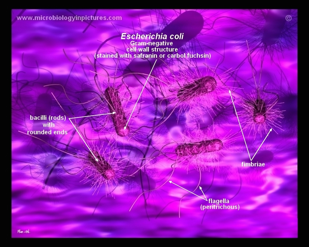
2 Morphology and Staining of Escherichia Coli E coli is Gramnegative straight rod, 13 µ x 0407 µ, arranged singly or in pairs (Fig 281) It is motile by peritrichous flagellae, though some strains are nonmotile Spores are not formed Capsules and fimbriae are found in some strains 3 Cultural Characteristics of Escherichia Coli4 Can a light microscope see bacteria?It is estimated that % of the human population are longterm carriers of S aureus
Many bacteria look like E coli when examined under the microscope (if not stained Enterobacteriaceae, Bacillus, cornyeformeSuppose your professor handed you a test tube with mL of an E Coli broth culture in it and told you to make a 10^2 dilution of the entire culture Explain how you would do this 100 fold dilution Dilute the 2 mL culture with 1980mL, to give a total solution of 100mLEndospore stained slide, with
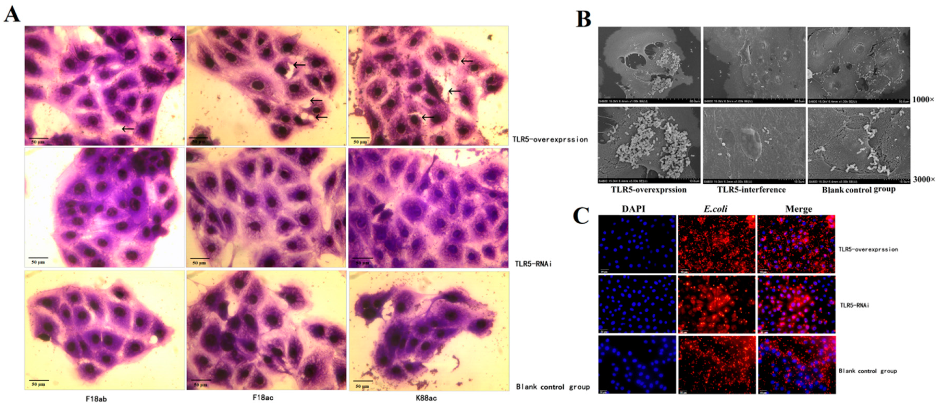


Animals Free Full Text Regulation And Molecular Mechanism Of Tlr5 On Resistance To Escherichia Coli F18 In Weaned Piglets Html
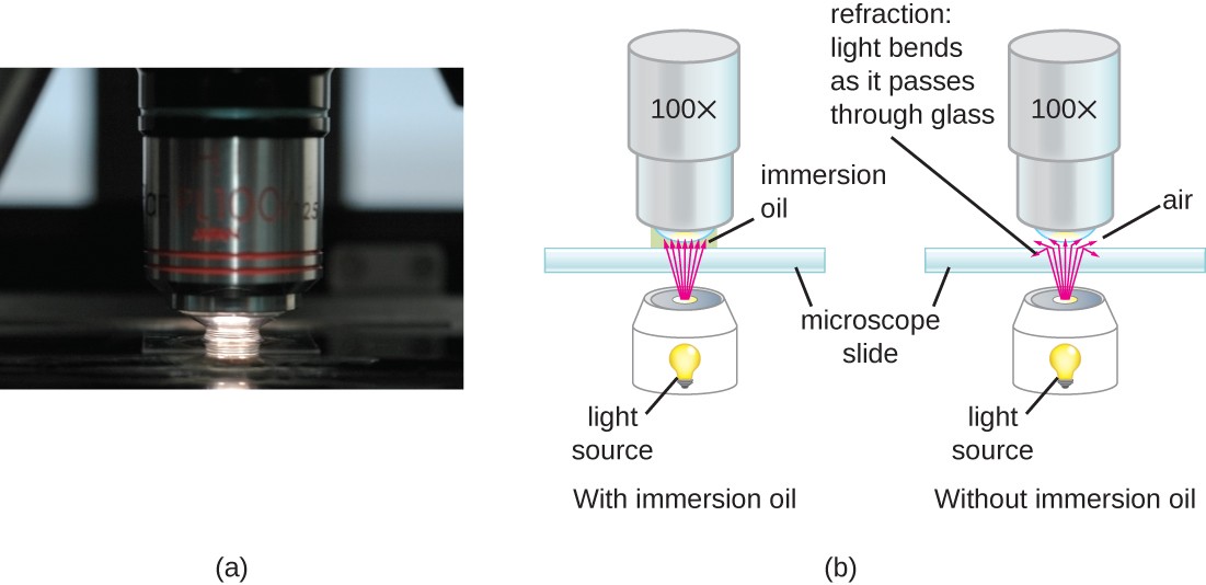


Instruments Of Microscopy Microbiology
Escherichia coli Four different strains of Escherichia coli on Endo agar with biochemical slope Glucose fermentation with gas production, urea and H 2 S negative, lactose positive (with exception of strain D "late lactose fermenter";1 Endospore stain of Bacillus subtilis showing both endospores (green) & vegetative cells (pink) @1000xTM;E coli stained gram negative under 1000x oil immersion
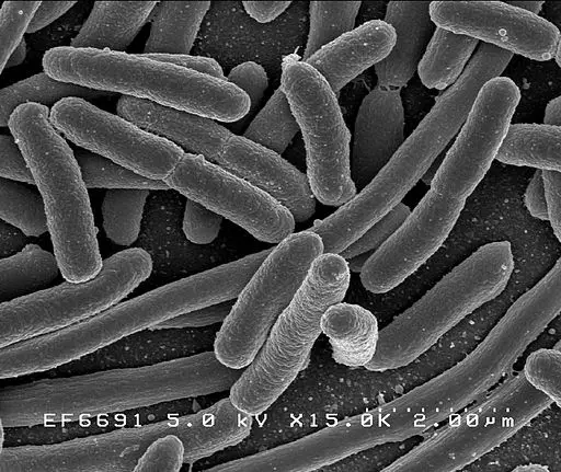


Observing Bacteria Under The Microscope Gram Stain Steps Rs Science
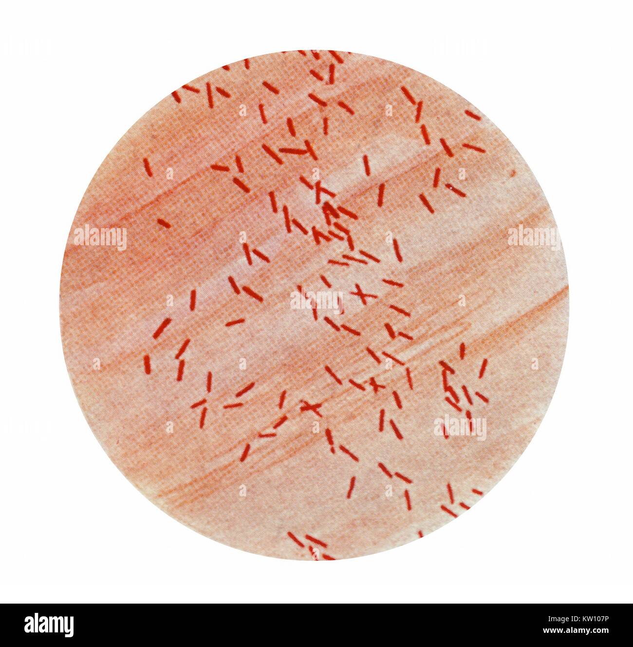


Gram Stain High Resolution Stock Photography And Images Alamy
This will give a total magnification of 1000X 4 Turn the light intensity control dial on the righthand side of the microscope to 6 Make sure the iris diaphragm lever in front under the stage is set approximately at 09, (toward the left side of the stage;Instead, their genetic material floats uncoveredThis will give a total magnification of 1000X 4 Turn the light intensity control dial on the righthand side of the microscope to 6 Make sure the iris diaphragm lever in front under the stage is set approximately at 09, (toward the left side of the stage;


Effect Of The Compound No On Cell Morphology Of E Coli Cells Download Scientific Diagram


Microbiology Lab Exercise 6 Acid Fast Staining Flashcards Cram Com
That should remain fully openExamples of Bacteria Under the Microscope Escherichia coli Escherichia coli (Ecoli) is a common gramnegative bacterial species that is often one of the first ones to be observed by students Most strains of Ecoli are harmless to humans, but some are pathogens and are responsible for gastrointestinal infections They are a bacillus shapedIn Figures below, various bacterial species including Micrococcus luteus, Staphylococcus epidermidis, Escherichia coli, Bacillus cereus, were observed under the microscope at 1000x magnification using gram staining which involves staining bacteria using crystal violet, and iodine as primary stain followed by decolouration with ethanol and staining with pink stain safranin


E Coli Stock Video Footage Royalty Free E Coli Videos Pond5


Chapter 3 Tools Of The Laboratory E Mail
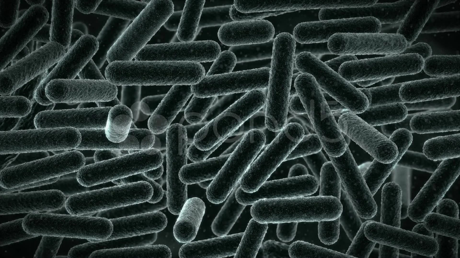
Escherichia coli, often abbreviated E coli, are rodshaped bacteria that tend to occur individually and in large clumps E coli are classified as facultative anaerobes, which means that they grow best when oxygen is present but are able to switch to nonoxygendependent chemical processes in the absence of oxygenE coli under the microscope Escherichia coli (E coli) is a bacterium commonly found in various ecosystems like land and water Most of the strains of E coli are harmless, but some strains are known to cause diarrhea and even UTIs E coli is commonly studied as they are considered as a standard for the study of different bacteriaAccording to the National Kidney Foundation, 80 to 90 percent of UTIs are caused by a bacteria called Escherichia coli For the most part, E coli lives harmlessly in your gut But it can cause


Micromorphology Slides Microbiology Resource Center Truckee Meadows Community College
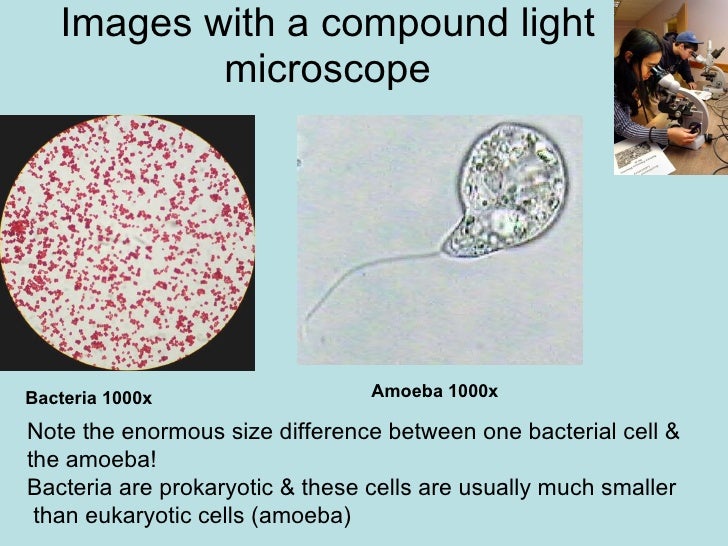


Chapter 3 Tools Of The Laboratory E Mail
Escherichia coli light microscopy Escherichia coli Magnification 1000×Yes, most of the bacteria range from 022 µm in diameter The length can range from 110 µm for filamentous or rodshaped bacteria The most wellknown bacteria E coli, their average size is ~15 µm in diameter and 26 µm in length As we talked above, you can see some bacteria in my cheek cells2 Morphology and Staining of Escherichia Coli E coli is Gramnegative straight rod, 13 µ x 0407 µ, arranged singly or in pairs (Fig 281) It is motile by peritrichous flagellae, though some strains are nonmotile Spores are not formed Capsules and fimbriae are found in some strains 3 Cultural Characteristics of Escherichia Coli


What Does An E Coli Bacteria Look Like Under A Microscope Quora
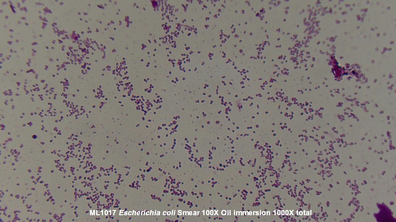


Slide Escherichia Coli
2 Negative endospore stain showing only vegetative cells @1000xTM;Escherichia coli, originally known as Bacterium coli commune, was identified in 15 by the German pediatrician, Theodor Escherich (14, 29) E coli is widely distributed in the intestine ofOscillatoria is about 7 µm in diameter The bacterium, Epulosiscium fishelsoni , can be seen with the naked eye (600 µm long by 80 µm in diameter)


Bacteria Under The Microscope E Coli And S Aureus Youtube


Capsule Staining Of E Coli E9 In The Absence And Presence Of Dpo42 Download Scientific Diagram
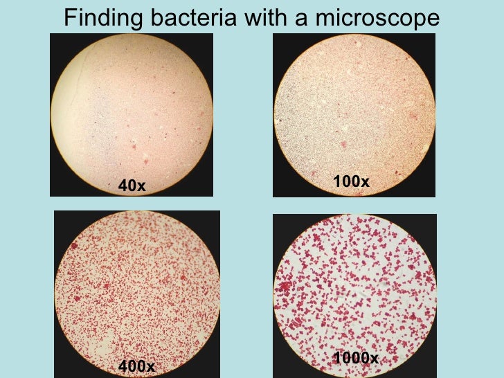
The total magnification of the microscope is calculated by multiplying the magnification of the objectives, with the magnification of the eyepiece Most educationalquality microscopes have a 10x (10power magnification) eyepiece and three objectives of 4x, 10x & 40x to provide magnification levels of 40x, 100x and 400xIs A 1 000x Zoom On A Microscope Enough To See Bacteria Cells Quora Microscope World Blog Bacillus Bacteria Under The Microscope Aquatic Microbial Ecology Igb E Coli Under The Microscope Types Techniques Gram Stain Gram Stained Bacterial Strain 68 Observed Under The Nikon EclipseE coli was discovered by Theodor Escherich in 15 after isolating it from the feces of newborns;
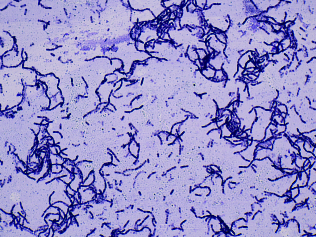


Solved In This Section You Will View A Random Set Of Micr Chegg Com
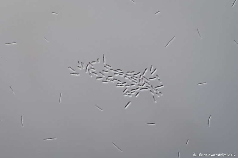


Usb Digital Microscope Microbehunter Com Microscopy Forum
3 Malachite green primary staining step of endopore stain with slide being heated over water bath;E Coli (Escherichia Coli) is a gramnegative, rodshaped bacterium Most E Coli strains are harmless, but some serotypes can cause food poisoning in their hosts The harmless strains are part of the normal flora of the gut Learn more about E Coli here Helicobacter Pylori The prepared microscope slide image of Helicobacter Pylori at left was captured at 400x magnificationThe most wellknown bacteria E coli, their average size is ~15 µm in diameter and 26 µm in length In this figure The size comparison between our hair (~ 60 µm) and E coli (~1 µm) Notice how small the bacteria are It requires 1000x magnification to see them well


My Microbe Assignment E Coli
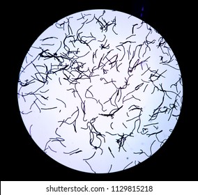


1000x Images Stock Photos Vectors Shutterstock
Escherichia coli (abbreviated as E coli) are bacteria found in the environment, foods, and intestines of people and animalsE coli are a large and diverse group of bacteria Although most strains of E coli are harmless, others can make you sick Some kinds of E coli can cause diarrhea, while others cause urinary tract infections, respiratory illness and pneumonia, and other illnessesRoyaltyfree stock photo ID Escherichia coli gram staining in compound microscope at 1000x zoomE coli stained gram negative under 1000x oil immersion


Morphology Of E Coli Cells Under Microscope At 100 Magnification Download Scientific Diagram


Biol 230 Lab Manual Lab 1
That should remain fully openE coli , a bacillus of about average size is 11 to 15 µm wide by to 60 µm long Spirochaetes occasionally reach 500 µm in length and the cyanobacterium;RESULTS After completing the experiment, it was concluded that M luteus is gram positive and E coli and S marcescens are gram negative As indicated in the student notes, the first slide that was prepared using aseptic technique and the Gram Staining method was a slide containing cultures of M luteus and E coli When examining this slide under the microscope at 1000X magnification, I observed some clusters and mostly doubles of coccishaped purple colonies


Bacterial Microscopy Streaked Images
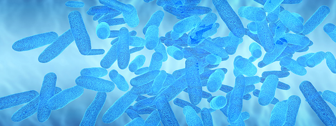


What Magnification Do I Need To See Bacteria Westlab
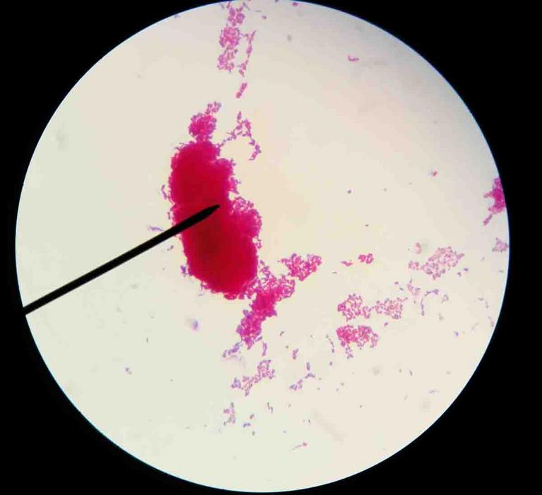
48/5 (428 Views 26 Votes) When properly Gramstained, Kocuria rhizophilia appears as purple spheres (Gram cocci) and Bacillus subtilis appears as light red rods (Gram– bacilli) at X magnification These two bacteria will be used as positive controls for comparison when Gramstaining a sample of E coli Click to see full answerSee Fig 6A) Do not close the field iris diaphragm ring on the light source;Escherichia coli Gram stained smear under microscopeRod shapedpink in colorthat's why Gram negative Bacilli#GramStain#GNB#GNR
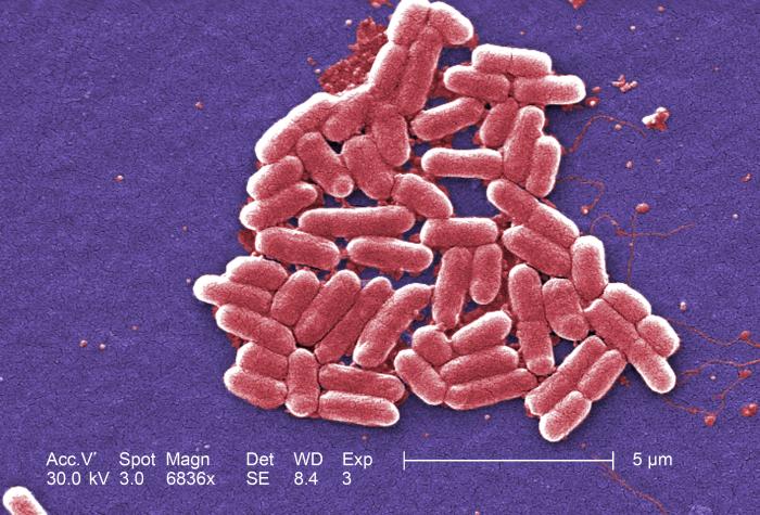


Details Public Health Image Library Phil


Is A 1 000x Zoom On A Microscope Enough To See Bacteria Cells Quora
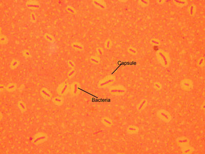
Cocci in grapelike clusters (Saureus) and bacilli(Ecoli) Clinical significance of Saureus Frequently found as part of the normal skin flora on the skin and nasal passages;Escherichia coli (/ ˌ ɛ ʃ ə ˈ r ɪ k i ə ˈ k oʊ l aɪ /), also known as E coli (/ ˌ iː ˈ k oʊ l aɪ /), is a Gramnegative, facultative anaerobic, rodshaped, coliform bacterium of the genus Escherichia that is commonly found in the lower intestine of warmblooded organisms (endotherms) Most E coli strains are harmless, but some serotypes (EPEC, ETEC etc) can cause serious foodE coli is Gramnegative straight rod, 13 µ x 0407 µ, arranged singly or in pairs (Fig 281) It is motile by peritrichous flagellae, though some strains are nonmotile Spores are not formed Capsules and fimbriae are found in some strains


Techniques Using A Microscope To Explore Fermented Foods Microbialfoods Org


Microscopy Aquarium Advice Aquarium Forum Community
Which bacteria look similar to E coli under 100X optical microscope?See Fig 6A) Do not close the field iris diaphragm ring on the light source;Escherichia coli bacteria on blood agar e coli under a microscope stock pictures, royaltyfree photos & images petri dishes with culture media for sarscov2 diagnostics, test coronavirus covid19, microbiological analysis e coli under a microscope stock pictures, royaltyfree photos & images
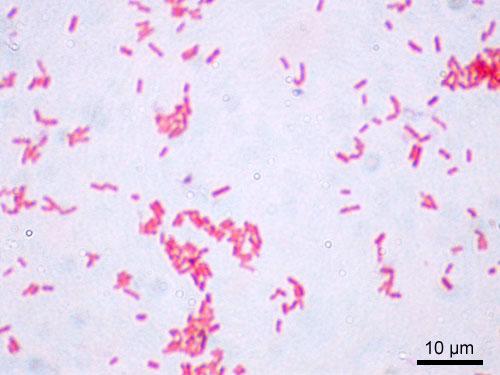


Laboratory Test 1 Flashcards Chegg Com


Can I See Bacteria With A 10x Microscope Quora
E Coli E Coli under the microscope at 400x E Coli (Escherichia Coli) is a gramnegative, rodshaped bacterium Most E Coli strains are harmless, but some serotypes can cause food poisoning in their hosts The harmless strains are part of the normal flora of the gut Learn more about E Coli here Helicobacter PyloriRESULTS After completing the experiment, it was concluded that M luteus is gram positive and E coli and S marcescens are gram negative As indicated in the student notes, the first slide that was prepared using aseptic technique and the Gram Staining method was a slide containing cultures of M luteus and E coli When examining this slide under the microscope at 1000X magnification, I observed some clusters and mostly doubles of coccishaped purple coloniesTo observe E coli with any detail, you will need to use the 100X lens, which is also known as an oil immersion lens This is the longest, most powerful and most expensive lens on the microscope, requiring extra care when using it As the name implies, the 100X lens is immersed in a drop of oil on the slide


Biol 230 Lab Manual Lab 1


Prey Range And Genome Evolution Of Halobacteriovorax Marinus Predatory Bacteria From An Estuary Biorxiv
Coliforms, E coli DOC modified mTEC prepared Agar 1 Method 67 Membrane Filtration Microscope, lowpower 1 Pipet(s) for dilution or for sample volumes less than 100 mL, if necessary 1 (1000x dilution) Mix well 8 If necessary, continue to dilute the sample 2 Coliforms, E coli, modified mTECThe length can range from 110 µm for filamentous or rodshaped bacteria The most wellknown bacteria E coli, their average size is ~15 µm in diameter and 26 µm in length In this figure The size comparison between our hair (~ 60 µm) and E coli (~1 µm) Notice how small the bacteria are It requires 1000x magnification to see them wellEscherichia coli or Ecoli is a gramnegative species of bacillus shaped bacteria that can be easily observed under a microscope, even for those with the untrained eye This bacteria has a fast growth rate, doubling every minutes, making them a common choice for bacterial related research purposes


Urine Sediment Of The Month Bacterial Variant Forms Renal Fellow Network


Pathogenic E Coli
The niche of E coli depends upon the availability of the nutrients within the intestine of host organisms;Since E coli are about 2 micrometres long, if you can't discriminate between two points that a four microns apart with your lens, you going to see a blurry smudge if you see anything at all It is routinely possible, using highend, oil immersion 100x lenses microscopes, to get down to 02 micrometre resolution, which is smaller than an E coli1 E coli stained with crystal violet @ 100xTM It is not possible to see individual bacteria at this magnification, but viewing at 100XTM is a necessary step in focusing to scope to see at higher magnifications 2 E coli stained with crystal violet @ 1000xTM
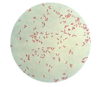


Pseudomonas Aeruginosa Microbewiki
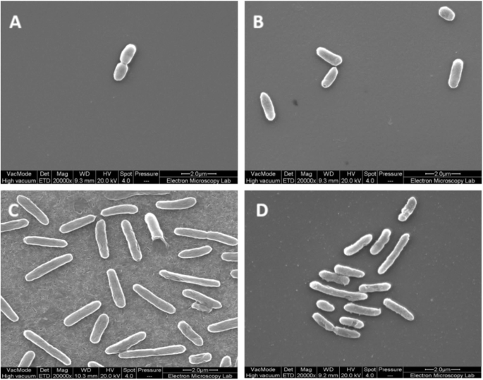


Thymol Tolerance In Escherichia Coli Induces Morphological Metabolic And Genetic Changes Bmc Microbiology Full Text
Biology Of E Coli E coli (Escherichia coli) are a small, Gramnegative species of bacteriaMost strains of E coli are rodshaped and measure about μm long and 0210 μm in diameterThey typically have a cell volume of 0607 μm, most of which is filled by the cytoplasm Since it is a prokaryote, E coli don't have nuclei;TELMU Microscope 40X1000X Dual Cordless LED Illumination Lab Compound Monocular Microscopes with Optical Glass Lenses & 10 Slides 2 Aspergillus WM 3 Three type of Smear 4 Coccus Smear 5 Root tip of plant LS 6 Agaricus sec 7 Spirogyia WM 8 Escherichia coli smear 9 Apical bud LS 10 Stem of monocotyledon TSE coli is described as a Gramnegative bacterium This is because they stain negative using the Gram stain The Gram stain is a differential technique that is commonly used for the purposes of classifying bacteria


Pbstatemicrobiology Licensed For Non Commercial Use Only Gram Stain


Asmscience Examination Of Gram Stains Of Urine
Flagellae (1000x) Flagellae are whiplike structures on a cell body that are used for locomotion, for feeding or other purposes Mouth Smear (400x) Human cheek cells divide about every 24 hours and are constantly shed from the body Cheek cells are often used to extract DNA for testing Escherichia Coli (400x)


How Bacillus Spores Look Like Under Light Microscope


Www Lycoming Edu Schemata Pdfs Fritz Bacterial growth Fall14 Pdf


Microscope World Blog Bacillus Subtilis Under The Microscope


Gram Stain


Gut Bacteria Escherichia Coli Under Microscope Youtube
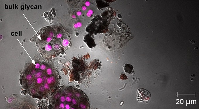


Instruments Of Microscopy Microbiology


3 Microscopic View 1000x Of Escherichia Coli Download Scientific Diagram


Evaluation Of Escherichia Coli Cells Damages Induced By Ultraviolet And Proton Beam Radiation
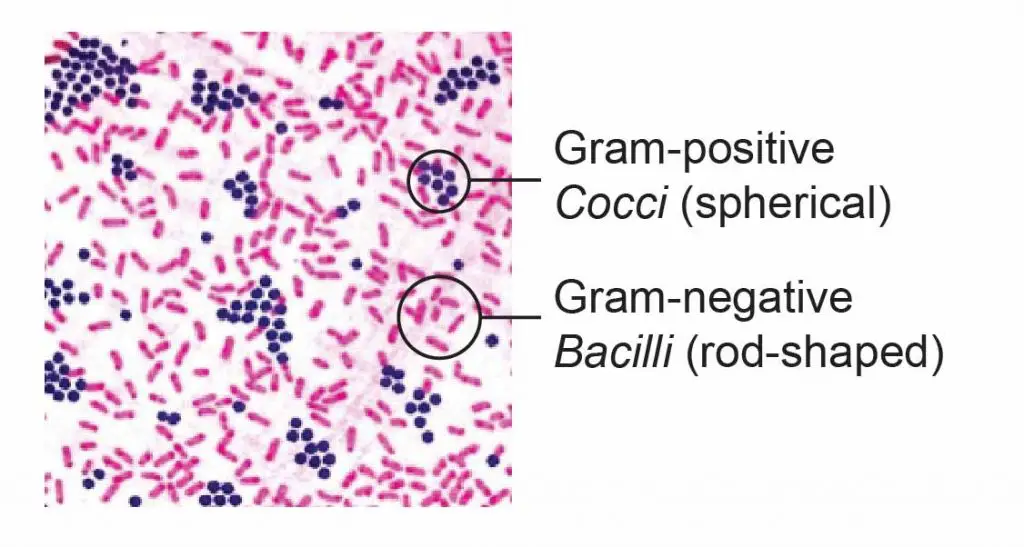


Observing Bacteria Under The Microscope Gram Stain Steps Rs Science


Gram Positive And Gram Negative Rods Microscopy Microbiology Microbiology Lab
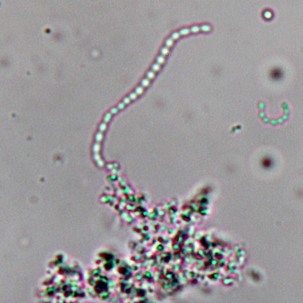


Observing Bacteria Under The Light Microscope Microbehunter Microscopy


Q Tbn And9gcswouuht13c4cxzkdgvwicdnmwhgkfjwlh40a Eerzzlpwjtmyt Usqp Cau
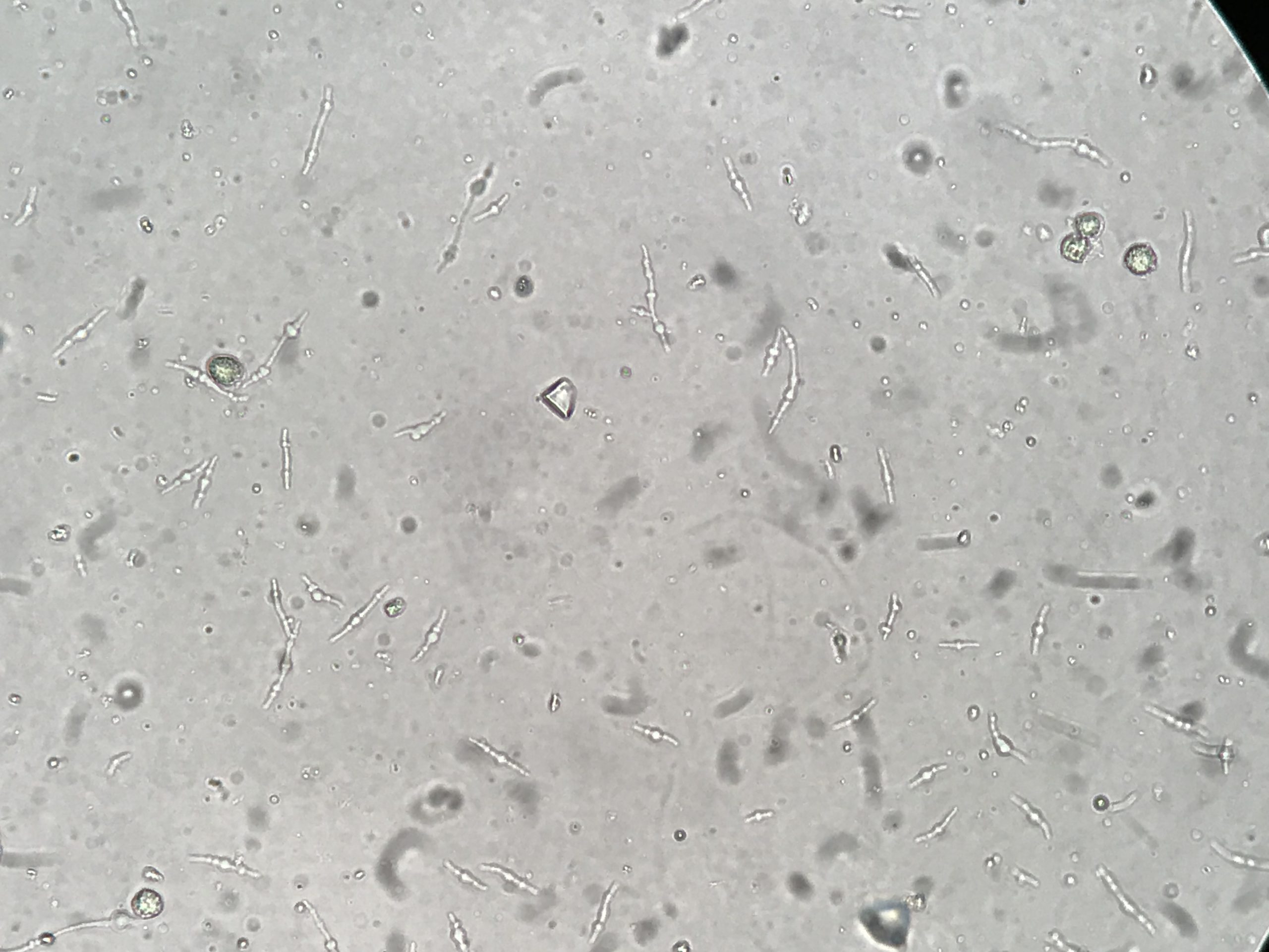


Urine Sediment Of The Month Bacterial Variant Forms Renal Fellow Network


Endospore Staining Principle Procedure And Results Learn Microbiology Online


Instruments Of Microscopy Microbiology


In Vitro Study Of The Activity Of Some Medicinal Plant Leaf Extracts On Urinary Tract Infection Causing Bacterial Pathogens Isolated From Indigenous People Of Bolangir District Odisha India Biorxiv


Bacterial Microscopy Streaked Images


Escherichia Coli Smear Gram Stain Prepared Microscope Slide 75 X 25mm Biology Microscopy Eisco Labs Amazon Co Uk Business Industry Science


Microscopy Gram Staining Microscope World Blog


Q Tbn And9gcr1gxralqmge Lwsjjzsouihvmyiv0ajl7a5fu8ma5voxpjs64l Usqp Cau


Escherichia Coli Light Microscopy


27 Earth Bacteria E Coli


Biol 230 Lab Manual Lab 1
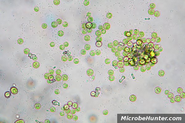


Observing Bacteria Under The Light Microscope Microbehunter Microscopy


E Coli Bacteria Under Microscope Page 1 Line 17qq Com
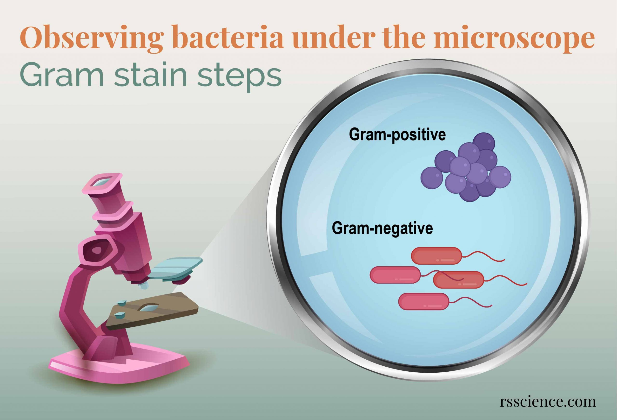


Observing Bacteria Under The Microscope Gram Stain Steps Rs Science


Bacterial Microscopy Streaked Images


How E Coli Bacteria Look Like


Escherichia Coli Bacteria E Coli Stock Footage Video 100 Royalty Free Shutterstock


44 Micro Ideas Microbiology Medical Laboratory Microbiology Lab


Gram Stain


Acid Fast Stain Free Microbiology Images From Science Prof Online


Avirulent Gram Negative Bacteria E Coli K 12 Or E Coli C Compared With Gram Positive Virulent Diplococcic Streptoccocus Pneumonia Insight Medical Publishing
.jpg)


Escherichia Coli 400x Escherichia Coli 400x Manufacturers Escherichia Coli 400x Suppliers Escherichia Coli 400x Exporters Escherichia Coli 400x In India


Lab 1
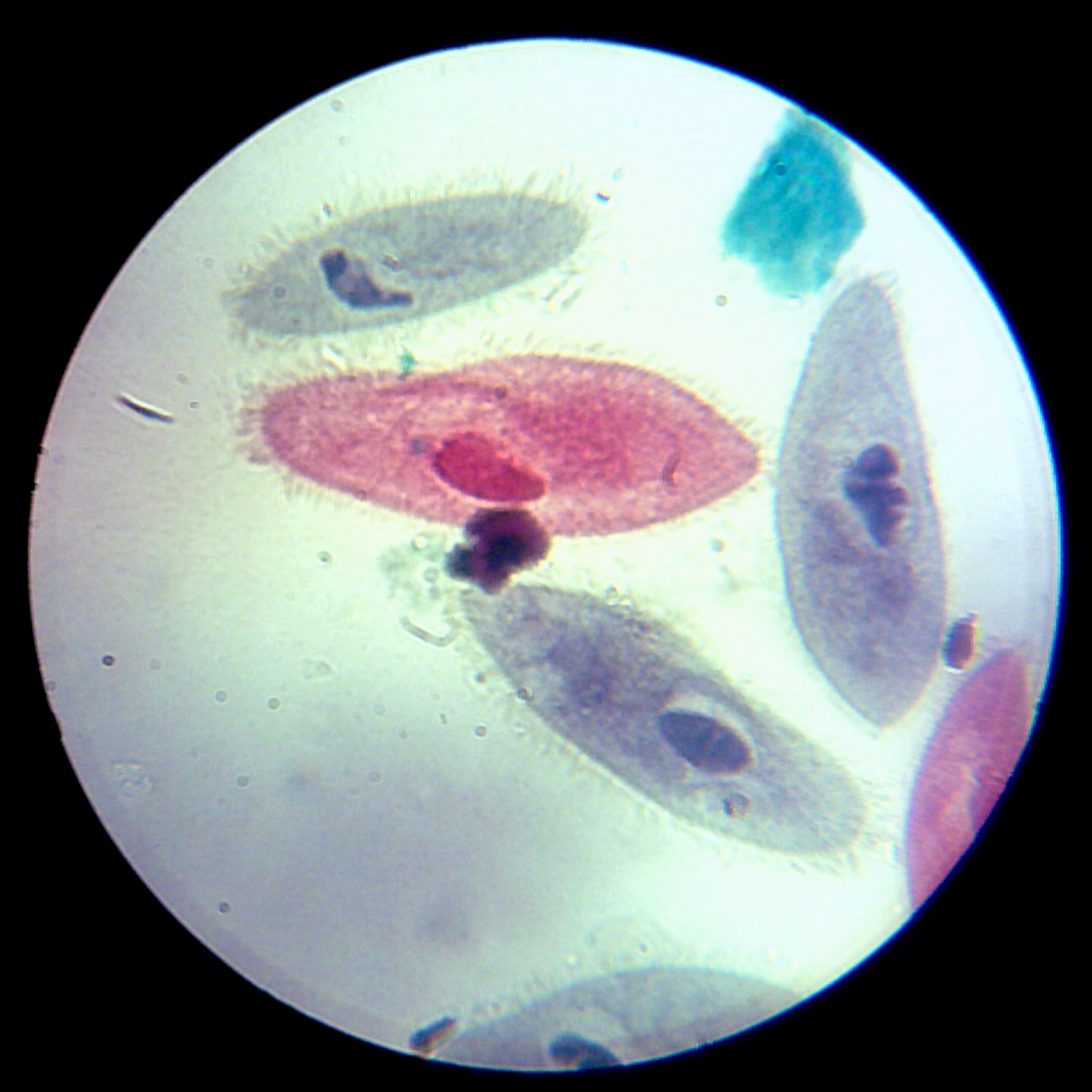


Under The Microscope Paramecium Office For Science And Society Mcgill University


Www Mccc Edu Hilkerd Documents Bio1lab3 Exp 4 Pdf


Http Coltonanderson1 Weebly Com Uploads 2 4 3 0 Manual Pdf


Direct E Coli Cell Count At Od600 Tip Biosystems


Www Mccc Edu Hilkerd Documents Bio1lab3 Exp 4 Pdf
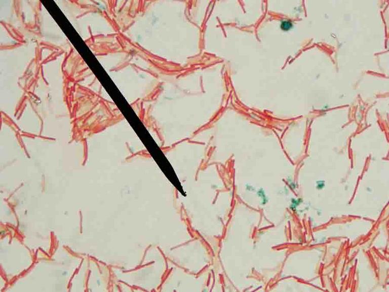


Endospore Bacterial Stain Microbiology Images Photographs From The Virtual Microbiology Classroom


Variation In Genome Content And Predatory Phenotypes Between ellovibrio Sp Nc01 Isolated From Soil And B Bacteriovorus Type Strain Hd100 Biorxiv
.jpg)


Bacteria 1000x Gram Stain Bacteria 1000x Gram Stain Manufacturers Bacteria 1000x Gram Stain Suppliers Bacteria 1000x Gram Stain Exporters Bacteria 1000x Gram Stain In India


Microscope World Blog Microscopy Gram Staining


E Coli Gram Stain Page 1 Line 17qq Com


Microscope Imaging Of Methylene Blue Stained E Coli Cells Harbouring Download Scientific Diagram


1000x Magnification Gram Drone Fest
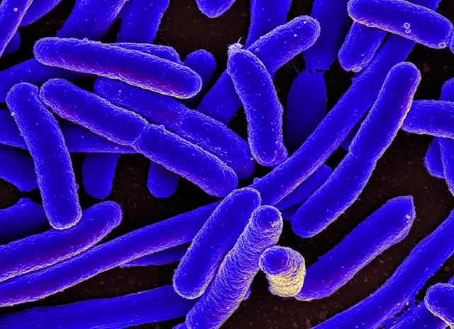


E Coli Under The Microscope Types Techniques Gram Stain Hanging Drop Method


Bacterial Microscopy Streaked Images


Cocci Bacteria Under Microscope Page 1 Line 17qq Com


Q Tbn And9gcqkye60ou Johpr02n Mbv1fferrjpdh Lnct7ymdf5qhyia1ld Usqp Cau


Q Tbn And9gcqkye60ou Johpr02n Mbv1fferrjpdh Lnct7ymdf5qhyia1ld Usqp Cau
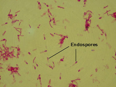


Micromorphology Slides Microbiology Resource Center Truckee Meadows Community College


Bacillus Subtilis Gram Stain Microbiology Bacillus Medical Laboratory Science


Coli Stock Footage Videos 180 Stock Videos


Escherichia Coli Bacteria E Coli Stock Footage Video 100 Royalty Free Shutterstock


Fimbria Bacteriology Wikipedia
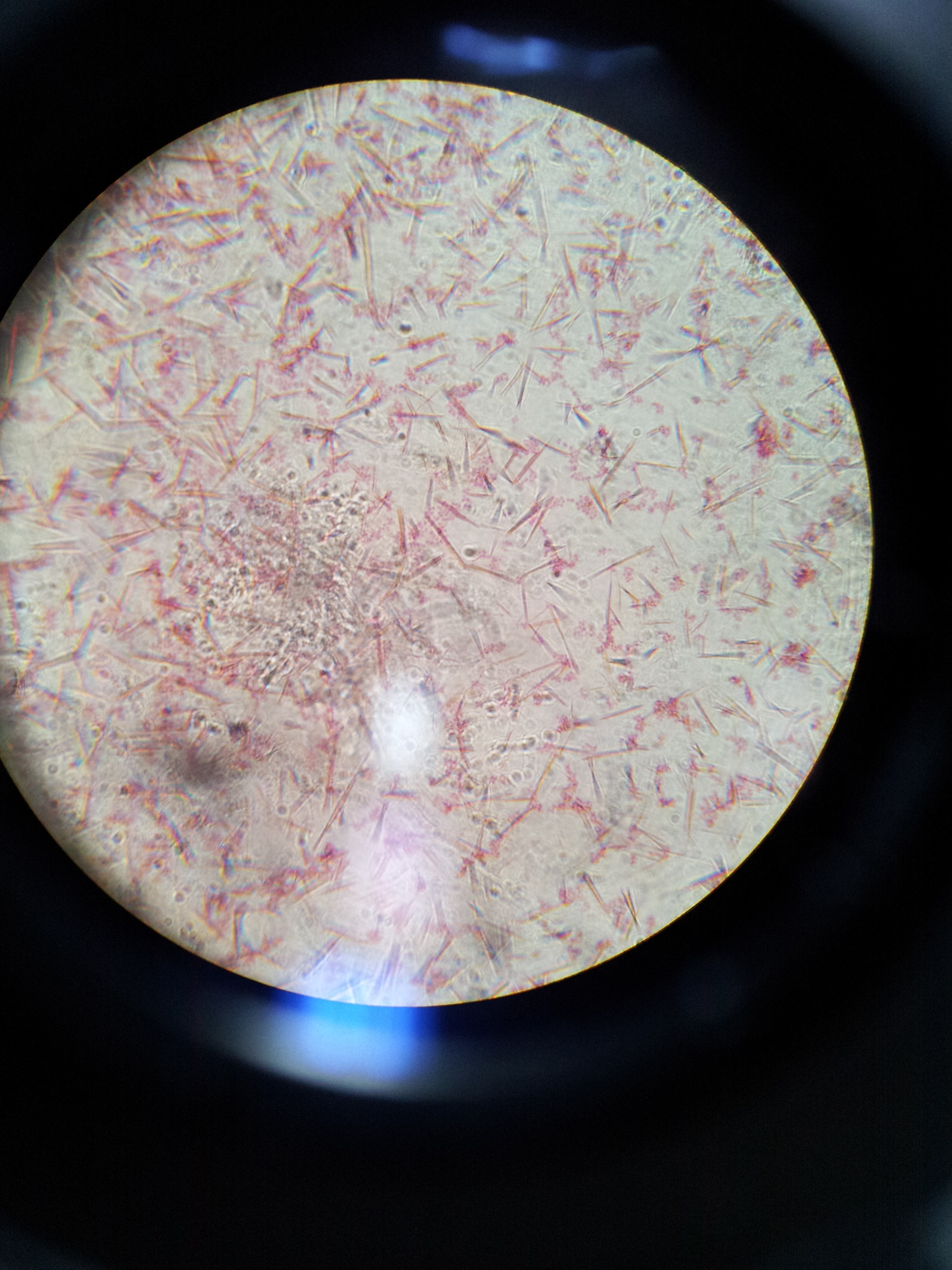


Lab 1 Principles And Use Of Microscope Ibg 102 Lab Reports
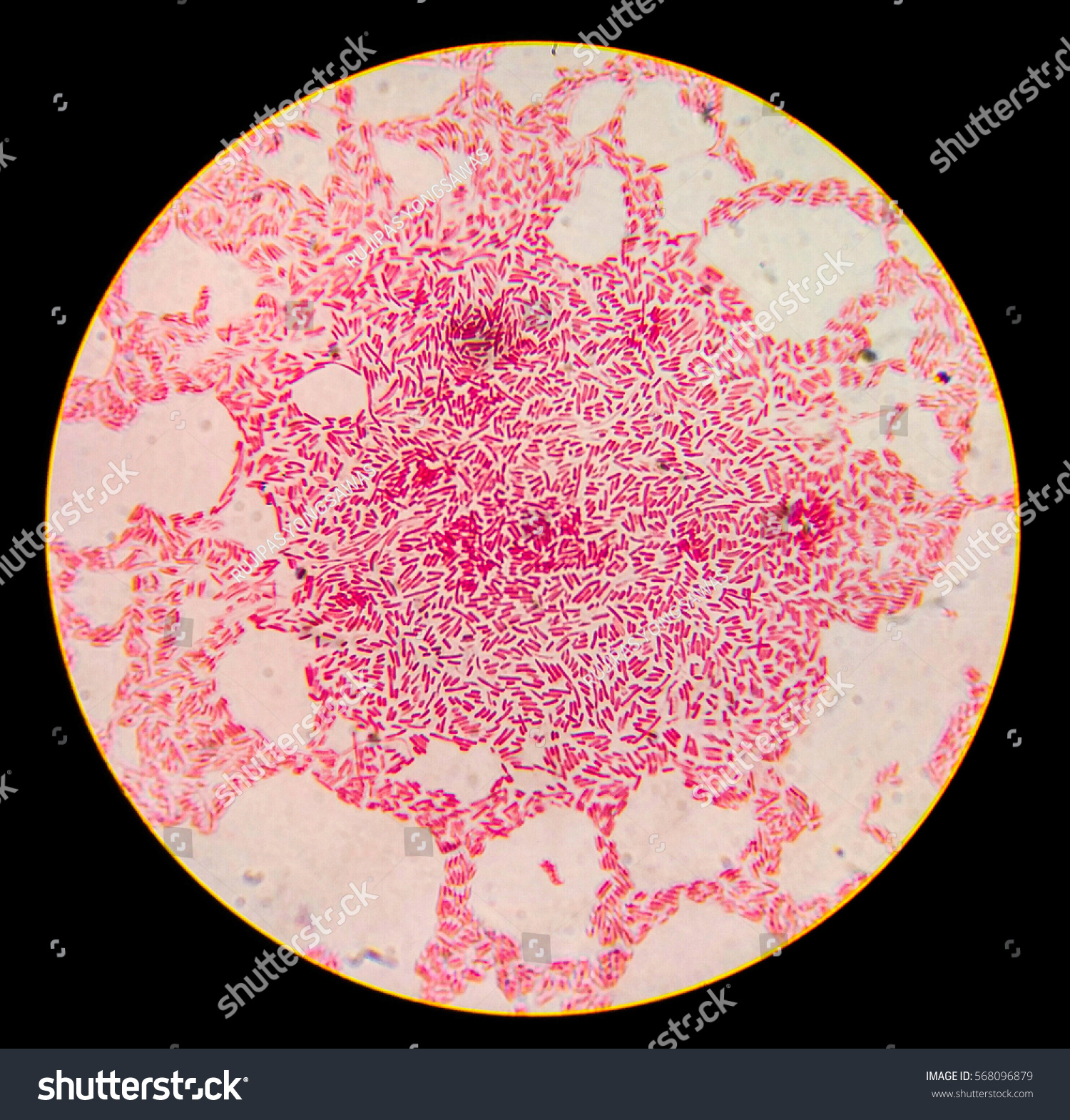


Escherichia Coli Gram Staining Compound Microscope Stock Photo Edit Now


White Blood Cells Crawling Under The Microscope 1000x Magnification Youtube


Gram Stain


Whats Your Microscopy Setup Advanced Mycology Shroomery Message Board
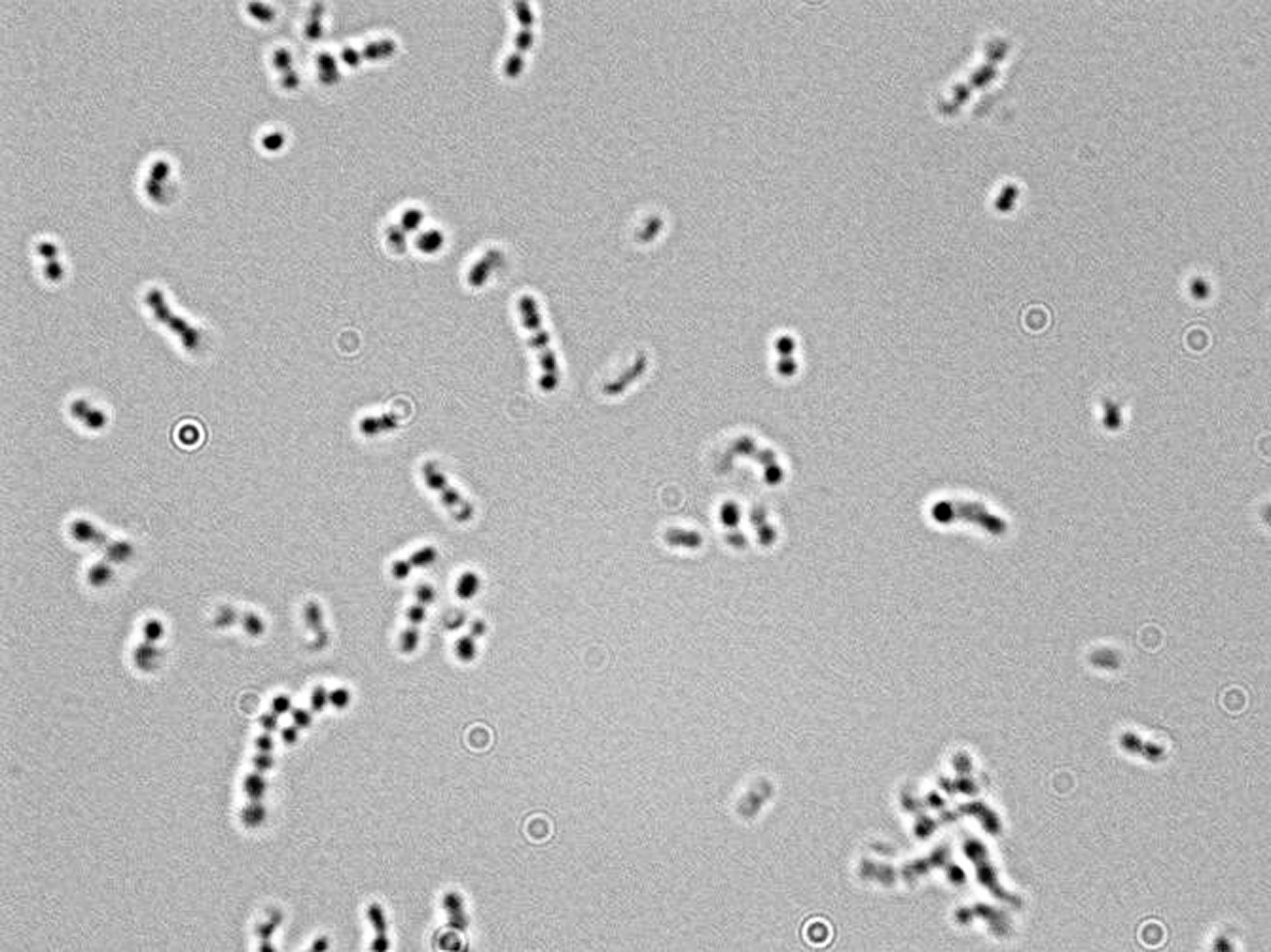


Microscopy For The Winery Viticulture And Enology


Pin On Microscopic Organisms


Escherichia Coli Bacteria E Coli Stock Footage Video 100 Royalty Free Shutterstock
/gram-positive-staphylococcus-aureus-bacteria-541802136-57979cca5f9b58461f26eccc.jpg)


Gram Stain Procedure In Microbiology


Royalty Free Escherichia Coli Bacteria E Coli Under Stock Video Imageric Com


Pathogenic E Coli


コメント
コメントを投稿